News

MCF is extending its Raman and PL capabilities to Deep UV with a new stand alone Renishaw system

  • Extremely high efficiency 250 mm focal length inVia Reflex spectrograph
  • Stand alone Renishaw Raman unit with solid-state Deep-UV laser (266 nm) and components
  • UV optics for high temperature and high power electronics.
  • Capability for Raman and PL spectroscopy from 200 nm – 1700 nm with automated mapping.
  • Andor InGaAs detector.
  • Ability to measure spectra of photonic materials deep into the UV range (e.g. AlxGa1-xN with up to 75% Al) including materials of Ultra-Wide Bandgap Initiatives.
  • Confocal Raman measurements with different Bright Field objective options
  • Different Grating options include 600 l/mm (NIR) & 3600 I/mm(UV)


Atomic Force Microscopy

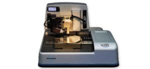
Bruker Dimension Icon Scanning Probe Microscopy
Performs all major SPM imaging like…

  • Peak Force Tapping
  • Contact Mode
  • Lateral Force Mode (LFM)
  • Electric Force Mode (EFM)
  • Magnetic Force Mode (MFM)
  • Fluid Imaging
  • Kelvin Probe Microscopy (KPFM)
  • Phase Imaging
  • Lift Mode
  • Force Spectroscopy
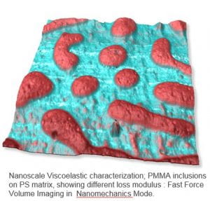
  • Force Volume
  • Surface Potential
  • Torsional Resonance (TR) Mode
  • Piezoresponse Microscopy

Samples up to 210 mm in dia/ 15 mm thick; Vertical range (Z) upto 13 microns; X,Y scan range -90 x 90 microns

Special Features

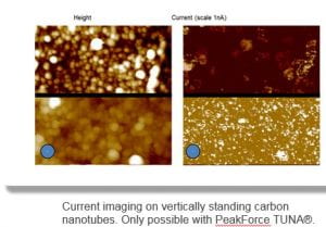
    • Can measure ultra low current between probe tip and sample using PeakForce TUNA Mode.
    • Can measure variation in conductivity and locate electrical defects of conductive samples using CAFM Mode.
    • New DataCube TUNA & SCM Modes provide Nanoelectrical & Nanomechanical spectra at every point.
    • Force Volume & Scan Asyst Probe Capabilities with user friendly software.

MCF May Image Contest!

The MCF May Image Contest is now live and you can submit your images here!

For information on the rules for submission, those can be seen here!

Also congratulations to the winners in April!
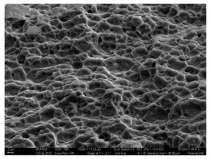
Sunflower – Lyu Zhiheng, Chen Ruhui (Xia – Chemistry)
Polymer Fingers – Liu Su, Tong Xin (Crittenden – Civil & Envir.)
Ultrathin GO membrane – Xin Tong, Liu Su (Crittenden – Civil & Envir.)
KHold Interface – Morris Satin (Rick Neu – ME)

MCF April Image Contest

The month of April is coming to a close, but it isn’t too late to submit an image to our image contest!

You can submit your image here!

If you would like to see all of our previous winners, you can see them here!

And congratulations to our winners in March 2019, Sang Yun Han, Erkul Karacauglu and Katherine Young!


Enter the EnvisioNano Image Contest! Deadline Dec 31, 2018!

EnvisioNano is a contest for undergraduate and graduate students conducting nanotechnology research in the United States and U.S. territories. Students should submit striking nanoscale images that demonstrate how beautiful the nanoscale can be alongside thoughtful, concise descriptions of the research behind the picture and how it may lead to nanotechnologies that benefit society. The goal is to envision where your research is headed and explain how “seeing” at the nanoscale is important to reaching that vision.  See the most recent winning image here!

For details and submission guidelines, please go here: https://www.nano.gov/EnvisioNano

December 2018 Image Contest is Live!

The December Image Contest is live and you can submit your images here!

If you have questions about the submission guidelines for the contest, those can be found on our website here.

And if you would like to see our previous winners (and congratulations to our winners in September and October), those images can be found here. The winner of the November contest will be posted by the end of this week.

If you have questions or concerns, please feel free to contact MCF Staff.

November 2018 Image Contest

The November Image Contest is live and you can submit your images here!

If you have questions about the submission guidelines for the contest, those can be found on our website here.

And if you would like to see our previous winners, those images can be found here.

If you have questions or concerns, please feel free to contact MCF Staff.

Webinar – How is Particle Size Measured

Masterclass
October 23
10:30 ET
The MCF will be showing this webinar on the monitor in Marcus at 10:30AM on October 23rd.
—-
There is a gamut of particle size distribution measurement techniques each with established history and advantages.

The killer word in particle size distribution is the last one (distribution) and measurement of broad distributions is the bane of all sizing techniques.

After a very short introduction to ‘how much sample should we measure to describe the distribution’, we’ll be running through the background of some of the more popular techniques (sieves, sedimentation, electrical-sensing zone) and will emphasize the popular light scattering techniques of laser diffraction and dynamic light scattering.

As an aside, we’ll be mentioning Small-Angle X-ray Scattering (SAXS).