Webinar on Advanced Battery Research

Battery research has rapidly grown in both scale and importance over the past decade. Join expert speakers who will be covering the latest research into optimizing existing energy storage solutions, developing novel technologies and deploying them in the real-world to meet our present and future energy demands.

Registration for the webinar can be found Advances in Battery Research 2024 Symposium | Technology Networks

If you find these links for webinars helpful, please contact David Tavakoli (atavakoli6 @ gatech.edu)

Renishaw Webinar on Particle Size Determination

The New Raman Renishaw Particle Analysis Software – Targeted Raman Data Collection

Wednesday, September 30th – 2:00pm (EST)

Renishaw’s upcoming Particle Analysis software enables targeted Raman data collection from optical image contrast. This approach ensures data is only collected from the areas of interest, making it a fast and automated method.

In this webinar, we will show how the diverse high performing optical contrast methods of the inVia Raman microscope can be used to quickly, easily, and automatically report particle identities and morphology together. Applied to a diverse range of applications, from microplastics to materials and forensics to pharmaceuticals, see how Particle Analysis can benefit your work.

This webinar will be broadcast in the MCF Lobby in the Marcus Nanotechnology Building.

Register for it here!


Rigaku Virtual Conference on XRD and XRF August 4-6!

Rigaku will be hosting a virtual conference on XRD and XRF this week that is free to register here!

Due to the COVID-19 induced cancellations of the Microscopy & MicroAnalysis, Denver X-ray, and American Crystallographic Association physical conferences this summer, Rigaku will be live webcasting a 3-day virtual Analytical X-ray Convention from our laboratory facility in Texas. The webcasts will take place Tuesday 8/4 – Thursday 8/6 and will feature live seminars on X-ray techniques and live instrument demonstrations.

Enjoy the presentations on Channel 1 (XRD), Channel 2 (XRF) and Channel 3 (X-ray Microscopy), and make sure to stop by our Concierge Booth on Channel 4 to say hello, live video chat, and participate in some fun events. We will be announcing upcoming Channel 4 events on the channel itself and via our Rigaku twitter feed (@rigaku, hashtag #RAXC2020), which you can view see on the right to keep up to date with what is going on.

Check the starting times for each day in the program, as different channels have different starting times. All four channels will be broadcast simultaneously and you can move among the four booths using the channel links.

Note that the three guest presentations—to be given by Rigaku sponsors—will all take place on channel 3. This has been updated on the program schedule.

Safety Videos Required for Returning to Campus

The MCF Staff look forward to welcoming our users back to campus and hope you are all safe and well.

From the office of the EVPR:

Research Ramp-Up Details


As part of a careful, gradual return to campus operations, research ramp-up activities are anticipated to begin as early as June 18. Only researchers and staff who must return to campus in order to carry out their job duties should return to work on campus. Supervisors will provide specific direction to their teams on when to return to in-person work. Anyone with questions about when or how they should start reporting to work in-person should, first, ask their supervisor.

Required: “Returning Safely to Your Lab” Video

If you have been identified by your supervisor to return to your lab, or if you have been working in one of the labs that has remained open, you will be required to watch the “Returning Safely to Your Lab” videos. The six-part module will include what you can expect, recommended safety precautions, cleaning and disinfection best practices, and proper use and disposal of PPE, among other helpful tips for best ensuring your safety and the safety of others.

Video Instructions:

  • Access the video by logging in at https://training.osp.gatech.edu/.
  • Click “My Online Classes” in left menu.
  • Select the first item: “Returning Safely to Your Lab.”
  • In “Available Courses” – click “Returning Safely to Your Lab.”
  • Click ‘Enroll Me.’
  • There are six video segments — when finished with each segment, go to the menu on the left side of the screen to start the next video.
  • After segment six, click Quiz in the menu and follow next steps to record that you have viewed the videos.


Webinar: Focus on Battery Research: Studying Battery Cathode Materials Using X-Ray Diffraction

Tuesday, April 22: 10:30-11:30AM.

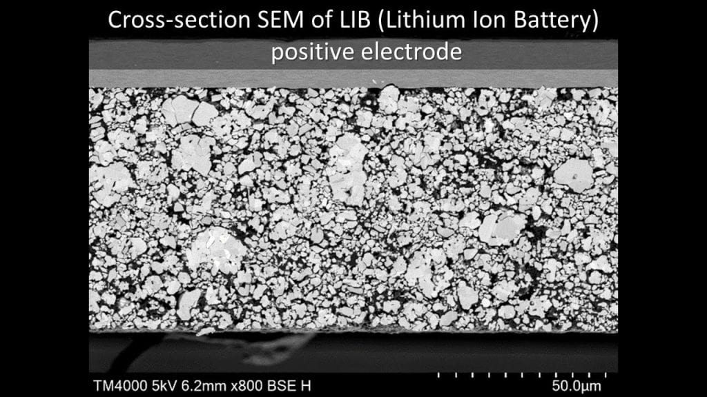
The cathode in a lithium-ion battery undergoes unique electrochemical reactions as lithium enters and leaves the atomic structure of the intercalated lithium compound. The intricacies of this reaction are one source of degradation and, therefore, an opportunity to improve performance. X-ray diffraction and scattering is well-suited to study these atomic phase changes, as well as a tool to understand and optimize the pathways that lithium uses to move through the cathode. However, studying battery materials requires special considerations that are different than the routine powder diffraction measurement.

This webinar, hosted by Malvern PANalytical will review the information that X-ray diffraction and scattering provides and discuss special considerations for experimental design such as selecting an X-ray tube, measurement geometry, and sample holder. We will then show examples of how these considerations are applied to cathode material analysis, including Rietveld refinement to quantify phase mixtures and atomic structure, pair distribution function analysis to examine local structural defects, and phase analysis of thick (10mm) commercial pouch cells, and in operando analysis of LFP based batteries to track phase changes during discharge and charging.

You can sign up for it here.

Speakers

Dr. Scott Speakman – Principal Scientist Malvern Panalytical
and Dr. Reeves-McLaren of The University of Sheffield

More information

– Who should attend?
Those working within the field of battery research or manufacturing or anyone interested in X-ray Diffraction of materials.
– What will you learn?
You will be educated on the X-ray diffraction and scattering application of lithium-ion batteries, including: Electrochemical reactions and atomic phase changes, with discussion on the special considerations needed for this application.


Webinar – XRD Masterclass 1 – Characterization Of Amorphous API

Webinar from Malvern PANalytical – XRD Masterclass 1 – Characterization Of Amorphous API 

Small molecule drug products often face multiple development challenges, common amongst which are those relating to solubility, stability and manufacturability.

The Developability Classification System (DCS) provides useful guidelines for selecting a formulation technology, based on assessment of the drug’s fundamental properties and dose expectations. APIs which fall under Class 1 (good solubility, good permeability) were discovered and delivered to the market early on.

Nowadays, the majority of small molecule drug candidates are poorly soluble and belong to Class 2 (a & b). For these molecules, solid-form screening and new formulation types are required to create competitive pharmaceutical products.

In the search for more soluble or bioavailable forms, different types of drug formulations are being  considered, including nanoparticles, amorphous solid dispersions, co-crystals and drug carrier systems. In this webinar, we’ll focus on amorphous formulations and address the following questions:

• Are amorphous compounds which are obtained in different experimental conditions the same?
• Are they free of nano-crystalline material and are they truly amorphous?
• What are the best ways of quantifying low and high amorphous content?
• How we can we use X-ray diffraction to answer these, and more, questions?

The MCF will be playing this webinar in the MCF Lobby but if you would like to register for this and watch it on your own computer, you can register here.

Date: February 20 2020

Time: 10:30 – 11:30
Webinar – Malvern PANalytical demonstration of Empyrean Series 3

The MCF will be showing a webinar on the new Malvern PANalytical Empyrean in the lobby tomorrow, Wednesday, December 18, 2019 from 10:30-11:30AM,

DEMO AT YOUR DESK – FLOOR STANDING XRD – THE EMPYREAN 3RD GENERATION

Join us for a demonstration of the New Empyrean 3rd generation X-ray diffractometer. Like no other system available, the Empyrean is designed for now, and for years to come. A fully automated series of 6 samples will be demonstrated using several different measurements types, including reflection geometry, SAXS, 2D transmission, texture, residual stress, thin film reflectivity, and grazing incidence XRD. The Optics enable the analyst a large variety of measurements without manual intervention. The predefined batch function with data collector has the programming power to switch between measurement types seamlessly. The world of materials science is constantly changing and the life of a high performance diffractometer like the Empyrean 3rd generation will deliver results that save time and effort,as well as, ensure accuracy of the experimental set up.


  • Who should attend?

– Anyone interested in XRD and the innovation of the floor standing X-ray diffraction platform


If you wish to watch this demonstration at your own desk or elsewhere, you can register for it here.

Webinar: Bringing EDS to life: sample considerations for biological element analysis – October 2nd!

Oxford Instruments webcast


The MCF will broadcast this webinar on the monitor in the lobby of the MCF in the Marcus Nanotechnology Building.

It will be streamed on Wednesday, October 2nd from 11:00AM-12:00PM. If you would like to watch it at your own computer, you can register for the webinar here!

Energy dispersive X-ray spectrometry (EDS) provides biologists with colourful element-based compositional information in addition to greyscale ultrastructural data produced with standard electron microscopy (EM), aiding the correct identification of structures and labels. A crucial aspect of all biological EM, including EDS, is the preparation of specimens with the aim of preserving and imaging samples as close to their living state as possible. The best option is freezing samples rapidly and imaging them in their frozen-hydrated state. However, the samples are sensitive to the electron beam requiring low dose imaging methods to avoid damage, and the low contrast makes identification of ultrastructure difficult. Chemical fixation allows the addition of contrasting agents and provides greater stability in samples, but prolonged preparation techniques may result in changes to ultrastructure and potential extraction of elements.

In this webcast, the speakers will discuss the challenges of biological EDS and provide information about sample preparation methods and imaging conditions in order to maximise results — from traditionally prepared samples to unstained specimens to cryo-electron microscopy of vitrified samples (CEMOVIS) — to identify and image cell ultrastructure in both transmission and scanning electron microscopy.