Measurement & Characterization December Demo Days in Pettit 160


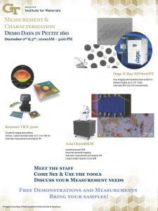
On Thursday and Friday, December 2nd & 3rd, the MCF in coordination with the IEN is hosting two days of measurement and characterization in the Pettit Microelectronics Building.

We plan to highlight previously available and new capabilities added over the last year for 2D, 3D, x-ray, and elemental measurements.

Please bring samples you are interested in measuring and staff will be happy to help you.

Download the flyer below, and we hope to see you there!

Where: Pettit 160
When: December 2nd & 3rd | 10:00AM – 5:00 PM

If you have questions or concerns, please feel free to contact the MCF Staff:

atavakoli6@gatech.edu, 770-689-6840

Pettit MCF Demo Day Advert Dec 2021

Webinar: Bringing EDS to life: sample considerations for biological element analysis – October 2nd!

Oxford Instruments webcast


The MCF will broadcast this webinar on the monitor in the lobby of the MCF in the Marcus Nanotechnology Building.

It will be streamed on Wednesday, October 2nd from 11:00AM-12:00PM. If you would like to watch it at your own computer, you can register for the webinar here!

Energy dispersive X-ray spectrometry (EDS) provides biologists with colourful element-based compositional information in addition to greyscale ultrastructural data produced with standard electron microscopy (EM), aiding the correct identification of structures and labels. A crucial aspect of all biological EM, including EDS, is the preparation of specimens with the aim of preserving and imaging samples as close to their living state as possible. The best option is freezing samples rapidly and imaging them in their frozen-hydrated state. However, the samples are sensitive to the electron beam requiring low dose imaging methods to avoid damage, and the low contrast makes identification of ultrastructure difficult. Chemical fixation allows the addition of contrasting agents and provides greater stability in samples, but prolonged preparation techniques may result in changes to ultrastructure and potential extraction of elements.

In this webcast, the speakers will discuss the challenges of biological EDS and provide information about sample preparation methods and imaging conditions in order to maximise results — from traditionally prepared samples to unstained specimens to cryo-electron microscopy of vitrified samples (CEMOVIS) — to identify and image cell ultrastructure in both transmission and scanning electron microscopy.