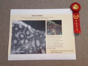
Congratulations to two of our staff members, Yolande Berta and Yong Ding on winning 2nd place for their submission of an “Owl” to the Ceramograph conference!
More information about the conference can be found here!
Congratulations to two of our staff members, Yolande Berta and Yong Ding on winning 2nd place for their submission of an “Owl” to the Ceramograph conference!
More information about the conference can be found here!
Every month the MCF hosts an image contest showing off the capabilities of our tools!
You can submit an image to be considered here!
https://gatech.infoready4.com/#competitionDetail/1795600
You can also see our previous winners here!
https://mcf.gatech.edu/monthly-image-contest-results/
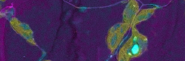
Congratulations to Yung Suk “Jeremy” Yoo for his image “Jellyfish” captured on the FEI Tecnai F30 and to Alexander Terwindt for his image of a Fly Eye captured on the Hitachi 8010!
The MCF will broadcast this webinar on the monitor in the lobby of the MCF in the Marcus Nanotechnology Building.
It will be streamed on Wednesday, October 2nd from 11:00AM-12:00PM. If you would like to watch it at your own computer, you can register for the webinar here!
—
Energy dispersive X-ray spectrometry (EDS) provides biologists with colourful element-based compositional information in addition to greyscale ultrastructural data produced with standard electron microscopy (EM), aiding the correct identification of structures and labels. A crucial aspect of all biological EM, including EDS, is the preparation of specimens with the aim of preserving and imaging samples as close to their living state as possible. The best option is freezing samples rapidly and imaging them in their frozen-hydrated state. However, the samples are sensitive to the electron beam requiring low dose imaging methods to avoid damage, and the low contrast makes identification of ultrastructure difficult. Chemical fixation allows the addition of contrasting agents and provides greater stability in samples, but prolonged preparation techniques may result in changes to ultrastructure and potential extraction of elements.
In this webcast, the speakers will discuss the challenges of biological EDS and provide information about sample preparation methods and imaging conditions in order to maximise results — from traditionally prepared samples to unstained specimens to cryo-electron microscopy of vitrified samples (CEMOVIS) — to identify and image cell ultrastructure in both transmission and scanning electron microscopy.
The MCF will be hosting a lecture on Atom Probe Tomography with scientists from Cameca on Wednesday, September 11, 2019 from 12:00 PM to 1:00 PM EDT .
This talk will cover APT operational theory, an introduction to sample prep and data reconstruction, and an overview of various applications. A commercial cryo-UHV solution for FIB-APT specimen transfer will also be presented.

If you would like to sign up, you can you do so here!
X-Ray Diffraction (XRD) is a powerful tool to look at crystals for characterizing microstructural and crystallographic properties of powders, thin films, fibers and other solid materials. The MCF has recently added another XRD, a Malvern PANalytical MPD to its capabilities. This XRD has a flat sample stage (default) for the analysis of powders and small solids, a non-ambient stage capable of running from -196C-450C, and a reflectivity stage. It should be on SUMS no later than 8/12/2019!
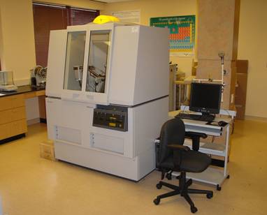
The Malvern PANalytical Empyrean in the MCF was featured prominently in a story recently posted online!
Many thanks to Neha Kondekar and Xenia Wirth as well as the MSE 2021 class for agreeing to participate!
If you would like to see the blog post, you can see it here!
—
The core facility for materials analysis at Georgia Tech is the IEN/IMat Materials Characterization Facility (MCF). The MCF is available to academic, industry and government users; it merges several labs on Georgia Tech’s campus and offers a variety of microscopy and characterization tools as well as skilled research staff to support research needs. Offering 24-hour a day shared-user access to the latest in imaging and analysis technology, and operated on a fee rate schedule, the MCF facility provides services for researchers including equipment training, remote sample prep and measurement, and imaging and analysis consultations.
MCF also happens to be where this top public research university and institute of technology houses their Malvern Panalytical Empyrean X-ray diffraction (XRD) instrument. The Empyrean XRD system generates X-rays, directs them toward a sample, and collects diffracted rays (the angle between the incident and the diffracted beam). Collected data are widely used for the identification of unknown crystalline materials (e.g. minerals, inorganic compounds), quantification of crystalline and amorphous materials, thin film thickness and structure, and many more applications. These applications are critical to studies in geology, environmental science, material science, engineering and biology.
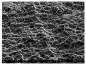
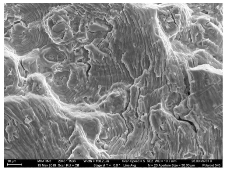
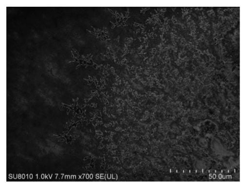
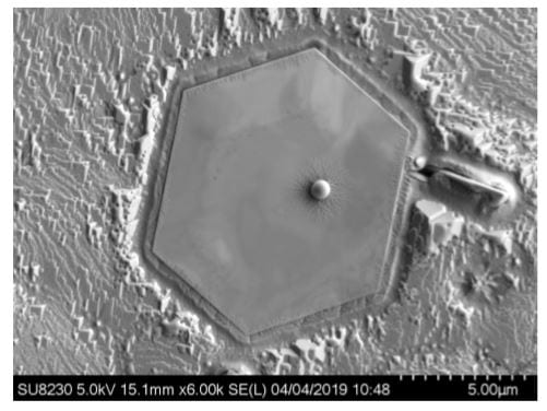
Congratulations to our image contest winners for May-June 2019!
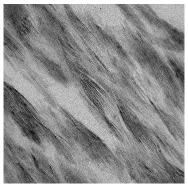
Morris Satin – Metallic Ripples
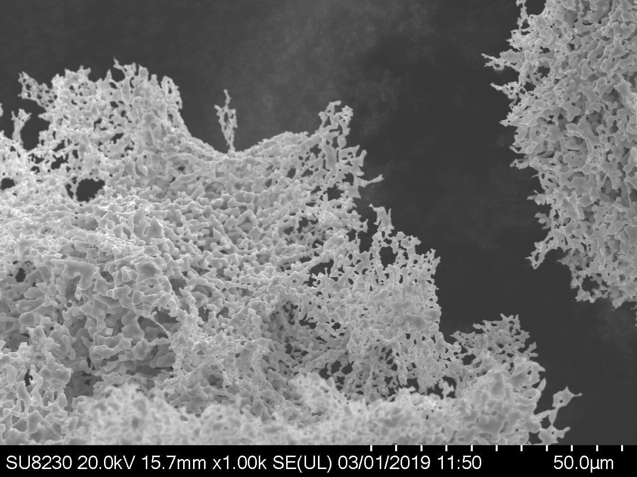
Jianshan Liao – Silicon Debris
Katie Young – Mo2C Flake on Copper
Zhiheng Lyu – “Sketches” – Copper Oxide Nanoparticles
You can see detailed images here as well as our previous winners!
The MCF May Image Contest is now live and you can submit your images here!
For information on the rules for submission, those can be seen here!
The month of April is coming to a close, but it isn’t too late to submit an image to our image contest!
You can submit your image here!
If you would like to see all of our previous winners, you can see them here!
And congratulations to our winners in March 2019, Sang Yun Han, Erkul Karacauglu and Katherine Young!
The MCF image competition for March is now open and you can submit your images here!
We want to show off your images and what our tools are capable of! If you would like to see the rules for submission you can see those here.
Morris Satin was the winner for January-February and you can see that image (and our previous images that won) here!