Webinar – Malvern PANalytical demonstration of Empyrean Series 3

The MCF will be showing a webinar on the new Malvern PANalytical Empyrean in the lobby tomorrow, Wednesday, December 18, 2019 from 10:30-11:30AM,

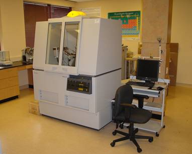
DEMO AT YOUR DESK – FLOOR STANDING XRD – THE EMPYREAN 3RD GENERATION

Join us for a demonstration of the New Empyrean 3rd generation X-ray diffractometer. Like no other system available, the Empyrean is designed for now, and for years to come. A fully automated series of 6 samples will be demonstrated using several different measurements types, including reflection geometry, SAXS, 2D transmission, texture, residual stress, thin film reflectivity, and grazing incidence XRD. The Optics enable the analyst a large variety of measurements without manual intervention. The predefined batch function with data collector has the programming power to switch between measurement types seamlessly. The world of materials science is constantly changing and the life of a high performance diffractometer like the Empyrean 3rd generation will deliver results that save time and effort,as well as, ensure accuracy of the experimental set up.


  • Who should attend?

– Anyone interested in XRD and the innovation of the floor standing X-ray diffraction platform


If you wish to watch this demonstration at your own desk or elsewhere, you can register for it here.

December Image Contest is now live!

The December Image Contest is live and you can submit your images here!

https://gatech.infoready4.com/#competitionDetail/1802759

We will be announcing our last months winners as well as our best images of the year next week!

Previous entries can be seen here!

If you are uncertain as to the rules of the submission process, those can be reviewed here.


New XANES/XAFS System Coming to the MCF!

The MCF is getting a new instrument capable of X-Ray Absorption Fine Structure (XAFS) and X-Ray Absorption Near Edge Structure (XANES).

This laboratory system for XAFS features a 1-2 kW conventional x-ray tube coupled to modern x-ray optics and detectors. It provides very rapid transmission-mode measurements suitable for research and development in electrical energy storage or catalysis while also giving extremely high throughput for general sample characterization or product testing. More details about the instrument can be found here from the instrument manufacturer.


Georgia Tech is hosting a workshop on XANES/XAFS techniques with an overview on Wednesday, December 11th and you can RSVP for that event here!


September MCF Image Contest is live!

Every month the MCF hosts an image contest showing off the capabilities of our tools!

You can submit an image to be considered here!

https://gatech.infoready4.com/#competitionDetail/1795600

You can also see our previous winners here!

https://mcf.gatech.edu/monthly-image-contest-results/

Congratulations to Yung Suk “Jeremy” Yoo for his image “Jellyfish” captured on the FEI Tecnai F30 and to Alexander Terwindt for his image of a Fly Eye captured on the Hitachi 8010!

Webinar: Bringing EDS to life: sample considerations for biological element analysis – October 2nd!

Oxford Instruments webcast


The MCF will broadcast this webinar on the monitor in the lobby of the MCF in the Marcus Nanotechnology Building.

It will be streamed on Wednesday, October 2nd from 11:00AM-12:00PM. If you would like to watch it at your own computer, you can register for the webinar here!

Energy dispersive X-ray spectrometry (EDS) provides biologists with colourful element-based compositional information in addition to greyscale ultrastructural data produced with standard electron microscopy (EM), aiding the correct identification of structures and labels. A crucial aspect of all biological EM, including EDS, is the preparation of specimens with the aim of preserving and imaging samples as close to their living state as possible. The best option is freezing samples rapidly and imaging them in their frozen-hydrated state. However, the samples are sensitive to the electron beam requiring low dose imaging methods to avoid damage, and the low contrast makes identification of ultrastructure difficult. Chemical fixation allows the addition of contrasting agents and provides greater stability in samples, but prolonged preparation techniques may result in changes to ultrastructure and potential extraction of elements.

In this webcast, the speakers will discuss the challenges of biological EDS and provide information about sample preparation methods and imaging conditions in order to maximise results — from traditionally prepared samples to unstained specimens to cryo-electron microscopy of vitrified samples (CEMOVIS) — to identify and image cell ultrastructure in both transmission and scanning electron microscopy.


Atom Probe Tomography for Atomic Scale Characterization and Biomaterials Analysis

The MCF will be hosting a lecture on Atom Probe Tomography with scientists from Cameca on Wednesday, September 11, 2019 from 12:00 PM to 1:00 PM EDT .

This talk will cover APT operational theory, an introduction to sample prep and data reconstruction, and an overview of various applications. A commercial cryo-UHV solution for FIB-APT specimen transfer will also be presented.

Atom Probe Tomography Schematic


If you would like to sign up, you can you do so here!


MCF is adding another XRD!

X-Ray Diffraction (XRD) is a powerful tool to look at crystals for characterizing microstructural and crystallographic properties of powders, thin films, fibers and other solid materials. The MCF has recently added another XRD, a Malvern PANalytical MPD to its capabilities. This XRD has a flat sample stage (default) for the analysis of powders and small solids, a non-ambient stage capable of running from -196C-450C, and a reflectivity stage. It should be on SUMS no later than 8/12/2019!

Image result for ford xrd gatech

A Day In The Life of an XRD!

The Malvern PANalytical Empyrean in the MCF was featured prominently in a story recently posted online!

Many thanks to Neha Kondekar and Xenia Wirth as well as the MSE 2021 class for agreeing to participate!

If you would like to see the blog post, you can see it here!

The core facility for materials analysis at Georgia Tech is the IEN/IMat Materials Characterization Facility (MCF). The MCF is available to academic, industry and government users; it merges several labs on Georgia Tech’s campus and offers a variety of microscopy and characterization tools as well as skilled research staff to support research needs. Offering 24-hour a day shared-user access to the latest in imaging and analysis technology, and operated on a fee rate schedule, the MCF facility provides services for researchers including equipment training, remote sample prep and measurement, and imaging and analysis consultations.

MCF also happens to be where this top public research university and institute of technology houses their Malvern Panalytical Empyrean X-ray diffraction (XRD) instrument. The Empyrean XRD system generates X-rays, directs them toward a sample, and collects diffracted rays (the angle between the incident and the diffracted beam). Collected data are widely used for the identification of unknown crystalline materials (e.g. minerals, inorganic compounds), quantification of crystalline and amorphous materials, thin film thickness and structure, and many more applications. These applications are critical to studies in geology, environmental science, material science, engineering and biology.


May-June 2019 Image Contest Winners!

Congratulations to our image contest winners for May-June 2019!

Morris Satin – Metallic Ripples

Jianshan Liao – Silicon Debris

Katie Young – Mo2C Flake on Copper

Zhiheng Lyu – “Sketches” – Copper Oxide Nanoparticles

You can see detailed images here as well as our previous winners!