Single point, 2D & 3D Chemical Imaging
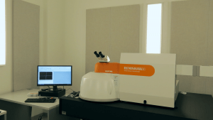 The Renishaw Qontor Raman Microscope uses a laser beam to excite the atoms of a material. Optical dispersion gratings and a sensitive camera permit the system to record the change in wavelength of the emitted spectrum vs the incident beam wavelength.
That spectrum of the light emitted as the atoms relax provides a fingerprint of the vibrations of the molecules in the sample.This fingerprint can used to identify the chemical bonds present in the sample, as well as molecular scale changes due to pressure, temperature, or other external stimuli.The data can be a single spectrum or a 2D map showing property variation at micron length scale.
To reserve time or request training on this tool, click here or on the image at left to go to the Raman tool page on SUMS.
 |
 |
 |
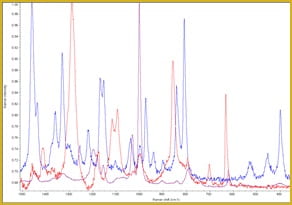
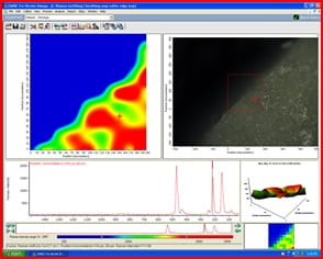
| Raman vibrational spectra of polystyrene (purple), PFA (blue), and PET(red), | Hyperspectral Raman map of a bulk sulfur sample. | Raman map (left) and optical image (right) of a ~5 μm graphene flake transistor with patterned electrodes |
Raman spectroscopy analyzes light that has been scattered inelastically so that it has changed wavelength. This wavelength change is a result of energy lost or gained after interaction with the vibrational states of the molecule that scatters the photon. By plotting the intensity of the scattered light versus the wavelength shift, one can obtain a spectral “fingerprint” for the molecules in a sample and their chemical and physical modifications.
Confocal microscopes focus light from a sample through a small pinhole to reject all light not in a very specific height plane – this allows them to create three-dimensional images of samples with high precision.
When this type of microscopy is combined with Raman spectroscopy the result is a Confocal Micro-Raman spectrometer (or Micro-Raman) system. This tool can generate 2D and even 3D chemical maps of solid, powder, liquid – and even gaseous – samples with submicron resolution.
[expand title="System Details:"]
The Raman system has two laser wavelengths for excitation – 488 nm and 785 nm
Stage:
– Can scan 110 mm in X and 75 mm in Y with a step size of 100 nm
– Can accommodate samples up to ~1cm thick
– Positioning accuracy to <1 μm, positioning repeatability to ~3 μm
– Integrated optical camera with zoom
– Manual and auto focus
Microscope/spectrometer:
– 5x, 20x, 50x long working distance (LWD), and 100x objectives
– Ability to select linear or circular polarization of incident and detected light
– Live-Track option to auto-focus on curved or rough surfaces in real time
– Incident laser intensity variable between 1E-6% and 100% of max power
Software:
– Automatic 1D line profiles, 1D depth profiles, and 2D & 3D maps
– Fast mapping option
– Library of Raman data for comparison/ID of materials
– Spectral correction for cosmic ray spikes,
– Automatic and/or Manual background correction, Peak ID, spectral math, peak fitting etc.
– Internal Si and Neon lamp standard for automatic calibration of system
[/expand]
Files:
Renishaw_Raman_SOP
WiRE 5 training script
introduction to Raman theory _Renishaw
Introduction to Confocal Raman microscopy_Renishaw
Introduction to Qontor Mapping_Renishaw
Renishaw Qontor-Standards,Alignment & Calibration