The X-ray Facility at Georgia Tech is split between two buildings.
1: In the Marcus Nanotechnology Building three Rigaku XRDs are installed. These instruments permit:
- Cryo measurements from 12K up to room temperature (Phenix Cryo stage)
- Non-ambient capillary measurements from ~90K to 600 deg. C (Anton-Paar TTK600 stage)
- Pair Distribution Function (PDF) measurements
- Auto-samplers for multi-sample measurements.
- Reactive in situ measurements up to 1000 deg. C can be performed under a variety of gasses. (ReactorX module from Rigaku)
- High-temp in situ measurements up to 2300 deg. C (HTK2000 hot stage from Anton-Paar)
- Ultra-fast Reciprocal Space Maps which should bring epitaxial measurement times down from hours to minutes
2: On the ground floor of the Paper Tricentennial Building are three Malvern PANalytical X-Ray Diffractometers (XRD’s). They are:
- The Alpha-1 which has an incident beam Johannsson monochromator and an X’Celerator solid-state detector,
- The Materials Research Diffractometer (MRD) The all-purpose MRD cradle is an Eulerian cradle designed to accommodate large samples and provide five motorized movements (phi, psi, X, Y, and Z). The mounting disk is parallel to the X-Y plane, Z direction is perpendicular to the mounting disk. The sample can be rotated about an axis perpendicular to its surface (phi movement) and/or around an axis parallel to its surface (psi movement).
- The Empyrean which has a high temperature furnace capable of reaching 1200 degrees Celsius for in-situ experiments, a 15-slot automatic sample changer, and Small Angle X-Ray Scattering (SAXS) capability.


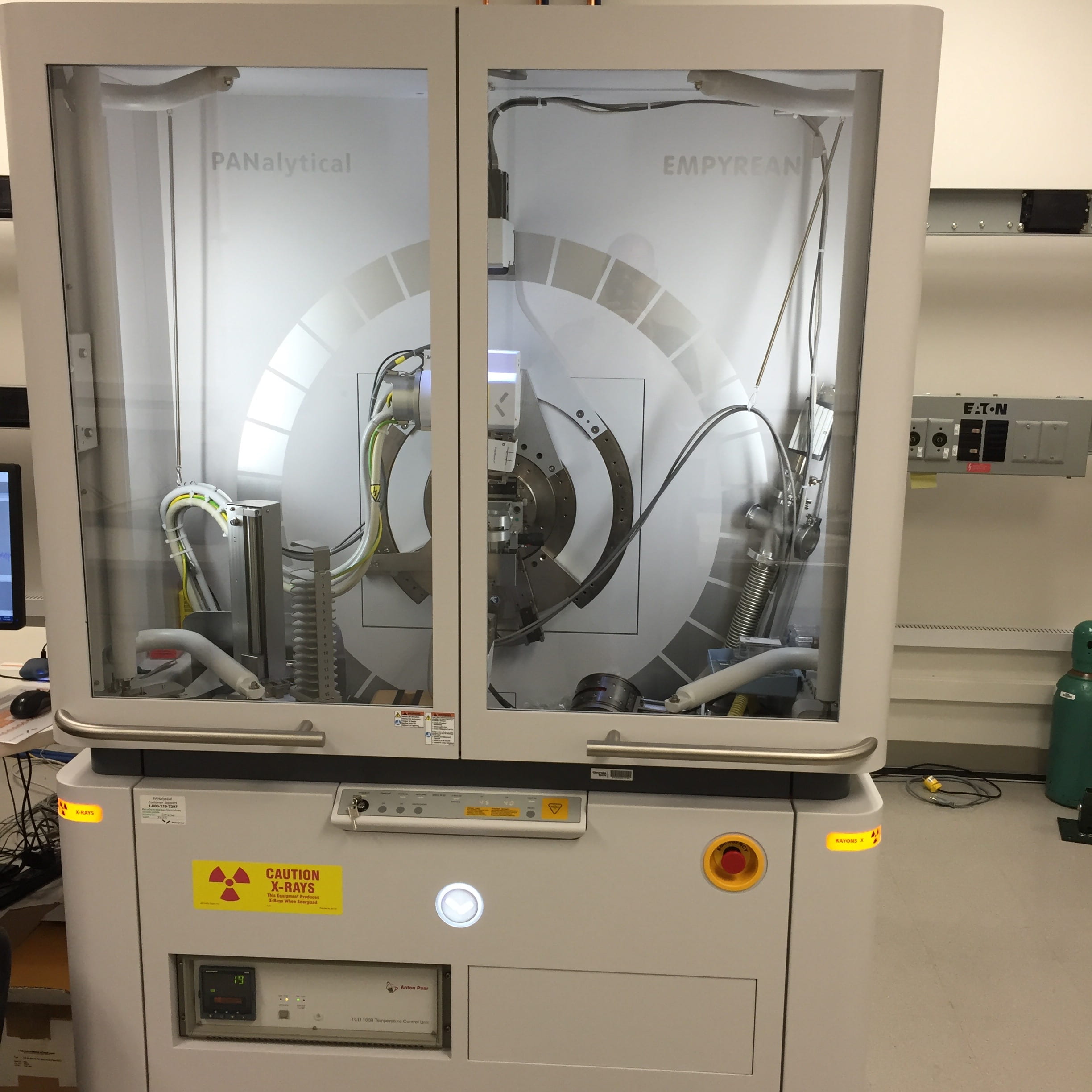
The functionality of these instruments is greatly multiplied by Malvern PANalytical’s revolutionary “PREFIX” optics system whereby instrument optics may be reconfigured with little more than the turn of a set screw, and an instrument may be changed from diverging beam optics to parallel beam optics in less than 5 minutes while still being completely aligned. The extensive range of measurements that can be performed using these instruments includes: Some Useful Links: Crystallography Frontiers: How Chemistry Reveals the Wonder of Everyday Materials This is intended as a (very) brief introduction to some of the common x-ray diffraction techniques used in materials characterization. It is designed for people who are novices in this field but are interested in using the techniques in their research. Extensive and authoritative discussions can be found in the numerous books and journal articles on this subject. Some references are listed below. X-rays primarily interact with electrons in atoms. When x-ray photons collide with electrons, some photons from the incident beam will be deflected away from the direction where they originally travel, much like billiard balls bouncing off one anther. If the wavelength of these scattered x-rays did not change (meaning that x-ray photons did not lose any energy), the process is called elastic scattering (Thompson Scattering) in that only momentum has been transferred in the scattering process. These are the x-rays that we measure in diffraction experiments, as the scattered x-rays carry information about the electron distribution in materials. On the other hand, in the inelastic scattering process (Compton Scattering), x-rays transfer some of their energy to the electrons and the scattered x-rays will have different wavelength than the incident x-rays. Diffracted waves from different atoms can interfere with each other and the resultant intensity distribution is strongly modulated by this interaction. If the atoms are arranged in a periodic fashion, as in crystals, the diffracted waves will consist of sharp interference maxima (peaks) with the same symmetry as in the distribution of atoms. Measuring the diffraction pattern therefore allows us to deduce the distribution of atoms in a material. The peaks in a x-ray diffraction pattern are directly related to the atomic distances. Let us consider an incident x-ray beam interacting with the atoms arranged in a periodic manner as shown in 2 dimensions in the following illustrations. The atoms, represented as green spheres in the graph, can be viewed as forming different sets of planes in the crystal (colored lines in graph on left). For a given set of lattice planes with an inter-plane distance of d, the condition for a diffraction (peak) to occur can be simply written as 2dsinθ = n λ which is known as Bragg’s law, after W.L. Bragg, who first proposed it. In the equation, λ is the wavelength of the x-ray, θ the scattering angle, and n an integer representing the order of the diffraction peak. Bragg’s Law is one of most important laws used for interpreting x-ray diffraction data. It is important to point out that although we have used atoms as scattering points in this example, Bragg’s Law applies to scattering centers consisting of any periodic distribution of electron density. In other words, the law holds true if the atoms are replaced by molecules or collections of molecules, such as colloids, polymers, proteins and virus particles. A small snapshot of some of the publications that have come out of our facility using the XRDs. Pharmaceutical crystallization in surface-modified nanocellulose organogels., If you would like training on any of these instruments and have a GTLogin; please log into SUMS, find the tool you would like to get trained on, and click on the request training button. That will start a forum conversation where we can discuss your sample and what kind of data you would like to generate, and we can determine the best instrument for your needs. If you have any questions, please contact: David Tavakoli (david.tavakoli@mse.gatech.edu)Functionality
Symmetry and Space Group Tutorial (Jerry P. Jasinski & Bruce M. Foxman)
International Union of Crystallography (IUCr) teaching pamphlets


Training